I did some research on hard coral diseases and their appearances. Heres what I found.
Black Band Disease: This disease is characterized by a "black mat", a few millimeters to centimeters (about 1/4 inch to 2 inches) wide, on the surface of coral tissue, moving across the surface of the skeleton, leaving behind bare white skeleton. The remaining coral tissue appears normal in color, morphology, and behavior.


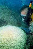
Bleaching: The loss of the microalgae or pigments from corals results in the white skeleton showing through the translucent tissue, thus it looks whitened or "bleached." (Note: The tissues of western tropical Atlantic massive starlet coral, Siderastrea siderea, can appear pinkish or bluish when bleaching occurs. Similarly, the finger coral, Porites porites, can appear bluish when bleached.) Bleaching can take several forms. The entire coral colony can become progressively lighter brown in color. Partial bleaching can occur, where some patches of tissue still retain the microalgae and photosynthetic pigments. At its most extreme, bleaching can result in all of the tissue appearing translucent white.
Patterns of bleaching on a colony can vary, with only the upper surface or lower surface of the colony being affected, or bleached tissue can appear as a circular patch or in the shape of a ring or a wedge. The pattern of bleaching may give important clues as to the cause of bleaching. The nature and extent of bleaching varies between individuals within a species and among species at the same location.
Dark Spots Disease: Dark purple to gray or brown patches of discolored tissue, often circular in shape but also occurring in irregular shapes and patterns, are scattered on the surface of the colony (bright purple patches have also been seen on a bleaching colony) or appear adjacent to the sediment/algal margin of a colony. Sediment can accumulate in the centers of these patches, with bare skeleton occasionally seen when the sediment is brushed off.

Rapid Wasting: Irregularly shaped, often large, patches of bare white skeleton appear on the surface of colonies of star coral (most commonly the columnar Montastraea annularis) and the brain coral Colpophyllia natans. The exposed skeletal surfaces are crumbly, often with the free edges of septa and pali missing or appearing to have eroded, with the result that the skeleton is depressed several millimeters to centimeters below the rest of the colony surface. The tissue margin can appear pale. The boundary between the skeleton and normal-appearing tissue is fairly sharp.
In early intermittent observations, it appeared that tissue loss advanced across the surface of the colony at the rate of several centimeters (2-3 inches) per day; usually stopping when the patch was from 5 to 50 centimeters (2 to 20 inches) across on star coral, or continuing until the tissue was completely lost from the colony on brain coral.

Red-Band Disease: As the name indicates, the "band" is a soft microbial mat that is brick red or dark brown, not black, in color and easily dislodged from the surface of the coral tissue. This disease affects hard star and brain corals (Diploria strigosa,Montastraea annularis, Montastraea cavernosa, Porites astreoides, Siderastrea radians, Colpophyllia natans) and sea fans in the Caribbean, and star, brain, and staghorn corals on the Great Barrier Reef.

White-Band Disease: Tissue peels or sloughs off the skeleton at a fairly uniform rate around the branch of the coral and progressing from the base of the branch towards the tip. On the staghorn coral (Acropora cervicornis), tissue loss may also occur in the middle of a branch. No consistent population of microorganisms has been found at the margin of the tissue loss.
All that can be seen with the naked eye is the tissue peeling off the bare white skeleton with occasional small bits of tissue remaining on the exposed skeleton. The rate of tissue loss is several millimeters (1/8 to 1/4 inch) per day. The bare skeleton is eventually colonized by filamentous algae, but the band of bare white skeleton that remains visible can be 5 to 10 centimeters (2 to 3 inches) wide. The tissue remaining on the branch shows no signs of pronounced bleaching, although affected colonies might appear slightly lighter in color overall. However, a variant of WBD, termed WBD Type II, has been found on staghorn colonies in the Bahamas. In this disease, a margin of bleached tissue appears before the tissue is lost.

White Plague: Tissue disappears from massive, encrusting, and branching species of corals at the rate of one or more centimeters (about 3/8 inch to 4 inches) per day (Florida Keys 1995), leaving behind bare white skeleton. The receding tissue appears as a sharp line at the skeletal interface. This rate is much faster than that previously reported for white plague, a few mm per day (Florida Keys 1977 and elsewhere). Many species have been affected by this kind of tissue loss.

White Pox: This disease is characterized by the appearance of irregularly-shaped patches of bare white skeleton. The patches can occur on the surface or undersides of branches. The tissue appears to peel off unevenly, but an even margin has also been found. The rate of tissue disappearance is unknown, but appears to be rapid, since the skeleton usually begins to be fouled by filamentous algae within a few days and the freshest patches can be several square inches (tens of square centimeters) in area but still have bare white skeleton. Gastropod predators have not been seen on most affected colonies of elkhorn, Acropora palmata.


Yellow-Blotch/Yellow-Band Disease: Yellow-blotch disease begins as an irregularly-shaped blotch of lightened yellow-colored tissue on the surface of the coral. As the disease progresses, the tissue in the center of the patch dies and the area fills with sediment and algae, resulting in a band of yellow tissue around the enlarging sediment patch (thus, it was first called yellow-band disease). The disease has been found most commonly on Montastraea faveolata, the massive keeled species, and less frequently on the columnar lobed star coral, Montastraea annularis.
In the disease affecting corals in the Arabian Gulf at Jebel Ali in Dubai, United Arab Emirates, the tissue is discolored yellowish and moves in a broad band with tissue loss and fouling of the skeleton with filamentous algae. Species affected are in the genera Acropora, Porites, Turbinaria, and Cyphastrea.


