disclaimer
The fate of the coral reefs lie in your hands.You decide.ToSave or don't save the reefs.(please click on the numbers to view the blog)profile
this blog was put up by a group of 3 from Kuo Chuan Presbyterian Primary School. :DWe are Lim Gun Hwi(the leader), Benjamin & Joseph Say.
Benjamin
Question: What is the importance of coral reefs?Answer:
Coral reefs are important for many different reasons:
The coral reef is "biologically diverse" that means there
are many different kinds of animals and plants that live theree
This is important, because just like the tropical rainforests, resources that haven't even been discovered yet, and unless we
Coral reefs may be the source of medicines, chemicals or other
these resources that we need.
Coral reefs are "biological
save these environments, we'll never get the chance to discove
rly productive" , which means there are lots of each thing that grows here. Many of the creatures that l orld!
Coral reefs are beautiful places, and provide income from
live here, like lobsters and fish, we depend on for food, even here in the Midwest , and all around the
w tourists to many otherwise poor countries around the world. Reefs are very stable, and provide a protective barrier around ct the reefs we have today, because they
can't be replaced.
many islands and coasts. Without the reefs these islands and coasts will erode away into the ocean. It is important to prot
e Reefs are formed very slowly by tiny animals called "coral". Each animal is tinier than your pinky nail, and grows very slowly, only about 1 centimeter a year. The reefs we have are big problems.
today were formed over 100s of thousands of years, and it would take just as long to grow back, if they grow back at all Oil drilling and erosion from developing coasts near reefs
Source :http://www.newton.dep.anl.gov/askasci/bio99/bio99276.htm
Hi, I would like you to watch the next few videos and just think about how beautiful these corals are. Just think if we continue polluting the sea and destroying the coral reefs.
There are many more other videos that show how beautiful these corals are. I hope that you will make a change and start helping to save the coral reefs. Everyone of us play a part in helping to save them.
Sorry for putting this post up so late. We should have placed this post on corals way earlier. So, just read on and you can find out more about those magnificent 'rainforest' of the sea.
Coral is a general term used to describe a group of cnidarians, which indicates the presence of skeletal material that is embedded in the living tissue or encloses the animal altogether. -National Oceanic and Atmospheric Administration, U.S. Dept. of Commerce. "Glossary of Coral Reef Terminology."
Corals themselves are tiny animals which belong to the group cnidaria (the "c" is silent). Other cnidarians include hydras, jellyfish, and sea anemones. Corals are sessile animals, meaning they are not mobile but stay fixed in one place.They feed by reaching out with tentacles to catch prey such as small fish and planktonic animals.
Corals are anthozoans, the largest class of organisms within the phylum Cnidaria. Comprising over 6,000 known species, anthozoans also include sea fans, sea pansies and anemones. Stony corals (scleractinians) make up the largest order of anthozoans, and are the group primarily responsible for laying the foundations of, and building up, reef structures. For the most part, scleractinians are colonial organisms composed of hundreds to hundreds of thousands of individuals, called polyps.
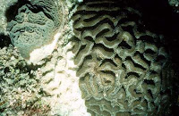
Corals live in colonies consisting of many individuals, each of which is called polyp. They secrete a hard calcium carbonate skeleton, which serves as a uniform base or substrate for the colony. The skeleton also provides protection, as the polyps can contract into the structure if predators approach. It is these hard skeletal structures that build up coral reefs over time. The calcium carbonate is secreted at the base of the polyps, so the living coral colony occurs at the surface of the skeletal structure, completely covering it. Calcium carbonate is continuously deposited by the living colony, adding to the size of the structure. Growth of these structures varies greatly, depending on the species of coral and environmental conditions-- ranging from 0.3 to 10 centimeters per year. Different species of coral build structures of various sizes and shapes ("brain corals," "fan corals," etc.), creating amazing diversity and complexity in the coral reef ecosystem. Various coral species tend to be segregated into characteristic zones on a reef, separated out by competition with other species and by environmental conditions.

Most corals are made up of hundreds of thousands individual polyps like this one. Many stony coral polyps range in size from one to three millimeters in diameter. Anatomically simple organisms, much of the polyp’s body is taken up by a stomach filled with digestive filaments. Open at only one end, the polyp takes in food and expels waste through its mouth. A ring of tentacles surrounding the mouth aids in capturing food, expelling waste and clearing away debris. Most food is captured with the help of special stinging cells called nematocysts which are inside the polyp' outer tissues, which is called the epidermis. Calcium carbonate is secreted by reef-building polyps and forms a protective cup called a calyx within which the polyps sits. The base of the calyx upon which the polyp sits is called the basal plate. The walls surrounding the calyx are called the theca. The coenosarc is a thin band of living tissue that connect individual polyps to one another and help make it a colonial organism.
As members of the phylum Cnidaria, corals have only a limited degree of organ development. Each polyp consists of three basic tissue layers: an outer epidermis, an inner layer of cells lining the gastrovascular cavity which acts as an internal space for digestion, and a layer called the mesoglea in between .
The diagram above shows the anatomy of a nematocyst cell and its “firing” sequence, from left to right. On the far left is a nematocyst inside its cellular capsule. The cell’s thread is coiled under pressure and wrapped around a stinging barb. When potential prey makes contact with the tentacles of a polyp, the nematocyst cell is stimulated. This causes a flap of tissue covering the nematocyst—the operculum—to fly open. The middle image shows the open operculum, the rapidly uncoiling thread and the emerging barb. On the far right is the fully extended cell. The barbs at the end of the nematocyst are designed to stick into the polyp’s victim and inject a poisonous liquid. When subdued, the polyp’s tentacles move the prey toward its mouth and the nematocysts recoil back into their capsules.
All coral polyps share two basic structural features with other members of their phylum. The first is a gastrovascular cavity that opens at only one end. At the opening to this cavity, commonly called the mouth, food is consumed and some waste products are expelled. A second feature all corals possess is a circle of tentacles, extensions of the body wall that surround the mouth. Tentacles help the coral to capture and ingest plankton for food, clear away debris from the mouth, and act as the animal’s primary means of defense.
While coral polyps have structurally simple body plans, they possess several distinctive cellular structures. One of these is called a cnidocyte—a type of cell unique to, and characteristic of, all cnidarians. Found throughout the tentacles and epidermis, cnidocytes contain organelles called cnidae, which include nematocysts, a type of stinging cell. Because nematocytes are capable of delivering powerful, often lethal toxins, they are essential to capturing prey, and facilitate coralline agonistic interactions.
Most corals, like other cnidarians, contain a symbiotic algae called zooxanthellae, within their gastrodermal cells. The coral provides the algae with a protected environment and the compounds necessary for photosynthesis. These include carbon dioxide, produced by coral respiration, and inorganic nutrients such as nitrates, and phosphates, which are metabolic waste products of the coral. In return, the algae produce oxygen and help the coral to remove wastes. Most importantly, they supply the coral with organic products of photosynthesis. These compounds, including glucose, glycerol, and amino acids, are utilized by the coral as building blocks in the manufacture of proteins, fats, and carbohydrates, as well as the synthesis of calcium carbonate (CaCO3). The mutual exchange of algal photosynthates and cnidarian metabolites is the key to the prodigious biological productivity and limestone-secreting capacity of reef building corals.
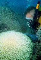
Zooxanthellae often are critical elements in the continuing health of reef-building corals. As much as 90% of the organic material they manufacture photosynthetically is transferred to the host coral tissue . If these algal cells are expelled by the polyps, which can occur if the colony undergoes prolonged physiological stress, the host may die shortly afterwards. The symbiotic zooxanthellae also confers its color to the polyp. If the zooxanthellae are expelled, the colony takes on a stark white appearance, which is commonly described as “coral bleaching”
Like global warming, ocean acidification is caused primarily by human emissions of carbon dioxide. In the atmosphere, that carbon helps to trap heat near the Earth's surface. In the oceans, it makes the water more acidic.
That increasingly acidic water threatens the viability of plankton, at the base of the food chain, as well as coral, because the acidic conditions prevent the formation of calcium carbonate shells. Some studies suggest many coral reefs will have died due to acidification by the end of this century. Coral reefs are the nurseries of the oceans, producing abundant fish of diverse species -- making them hotspots for tourism, and important areas for the fishing industry. Scientists at the International Coral Reef Symposium this July declared ocean acidification the largest and most significant threat to oceans, a significant statement, considering the vast body of evidence that overfishing and other forms of pollution are taking a massive toll on ocean life.
“Coral reefs are at the heart of our tropics, and millions of people around the world depend on these systems for their livelihoods. Without urgent action to limit carbon dioxide emissions and improve management of marine protected areas, even vast treasured reefs like the Great Barrier Reef and Northwestern Hawaiian Islands will become wastelands of dead coral,” said Lynne Hale, director of The Nature Conservancy’s Marine Initiative.
The scientists, convened by The Nature Conservancy in August, have agreed to the "Honolulu Declaration on Ocean Acidification and Reef Management." Here are some of its key elements:
Limit fossil fuel emissions
Build the resilience of tropical marine ecosystems and communities to maximize their ability to resist and recover from climate change impacts, including ocean acidification
Create new marine reserves that include those reefs less vulnerable to acidification, and consider climate change as a factor in managing existing reserves
Increase money spent on science and develop an international network to monitor and study acidification
Decrease other stresses to coral reefs, such as agricultural runoff and overfishing
I did some research on hard coral diseases and their appearances. Heres what I found.
Black Band Disease: This disease is characterized by a "black mat", a few millimeters to centimeters (about 1/4 inch to 2 inches) wide, on the surface of coral tissue, moving across the surface of the skeleton, leaving behind bare white skeleton. The remaining coral tissue appears normal in color, morphology, and behavior.


Bleaching: The loss of the microalgae or pigments from corals results in the white skeleton showing through the translucent tissue, thus it looks whitened or "bleached." (Note: The tissues of western tropical Atlantic massive starlet coral, Siderastrea siderea, can appear pinkish or bluish when bleaching occurs. Similarly, the finger coral, Porites porites, can appear bluish when bleached.) Bleaching can take several forms. The entire coral colony can become progressively lighter brown in color. Partial bleaching can occur, where some patches of tissue still retain the microalgae and photosynthetic pigments. At its most extreme, bleaching can result in all of the tissue appearing translucent white.
Patterns of bleaching on a colony can vary, with only the upper surface or lower surface of the colony being affected, or bleached tissue can appear as a circular patch or in the shape of a ring or a wedge. The pattern of bleaching may give important clues as to the cause of bleaching. The nature and extent of bleaching varies between individuals within a species and among species at the same location.
Dark Spots Disease: Dark purple to gray or brown patches of discolored tissue, often circular in shape but also occurring in irregular shapes and patterns, are scattered on the surface of the colony (bright purple patches have also been seen on a bleaching colony) or appear adjacent to the sediment/algal margin of a colony. Sediment can accumulate in the centers of these patches, with bare skeleton occasionally seen when the sediment is brushed off.

Rapid Wasting: Irregularly shaped, often large, patches of bare white skeleton appear on the surface of colonies of star coral (most commonly the columnar Montastraea annularis) and the brain coral Colpophyllia natans. The exposed skeletal surfaces are crumbly, often with the free edges of septa and pali missing or appearing to have eroded, with the result that the skeleton is depressed several millimeters to centimeters below the rest of the colony surface. The tissue margin can appear pale. The boundary between the skeleton and normal-appearing tissue is fairly sharp.
In early intermittent observations, it appeared that tissue loss advanced across the surface of the colony at the rate of several centimeters (2-3 inches) per day; usually stopping when the patch was from 5 to 50 centimeters (2 to 20 inches) across on star coral, or continuing until the tissue was completely lost from the colony on brain coral.

Red-Band Disease: As the name indicates, the "band" is a soft microbial mat that is brick red or dark brown, not black, in color and easily dislodged from the surface of the coral tissue. This disease affects hard star and brain corals (Diploria strigosa,Montastraea annularis, Montastraea cavernosa, Porites astreoides, Siderastrea radians, Colpophyllia natans) and sea fans in the Caribbean, and star, brain, and staghorn corals on the Great Barrier Reef.

White-Band Disease: Tissue peels or sloughs off the skeleton at a fairly uniform rate around the branch of the coral and progressing from the base of the branch towards the tip. On the staghorn coral (Acropora cervicornis), tissue loss may also occur in the middle of a branch. No consistent population of microorganisms has been found at the margin of the tissue loss.
All that can be seen with the naked eye is the tissue peeling off the bare white skeleton with occasional small bits of tissue remaining on the exposed skeleton. The rate of tissue loss is several millimeters (1/8 to 1/4 inch) per day. The bare skeleton is eventually colonized by filamentous algae, but the band of bare white skeleton that remains visible can be 5 to 10 centimeters (2 to 3 inches) wide. The tissue remaining on the branch shows no signs of pronounced bleaching, although affected colonies might appear slightly lighter in color overall. However, a variant of WBD, termed WBD Type II, has been found on staghorn colonies in the Bahamas. In this disease, a margin of bleached tissue appears before the tissue is lost.

White Plague: Tissue disappears from massive, encrusting, and branching species of corals at the rate of one or more centimeters (about 3/8 inch to 4 inches) per day (Florida Keys 1995), leaving behind bare white skeleton. The receding tissue appears as a sharp line at the skeletal interface. This rate is much faster than that previously reported for white plague, a few mm per day (Florida Keys 1977 and elsewhere). Many species have been affected by this kind of tissue loss.

White Pox: This disease is characterized by the appearance of irregularly-shaped patches of bare white skeleton. The patches can occur on the surface or undersides of branches. The tissue appears to peel off unevenly, but an even margin has also been found. The rate of tissue disappearance is unknown, but appears to be rapid, since the skeleton usually begins to be fouled by filamentous algae within a few days and the freshest patches can be several square inches (tens of square centimeters) in area but still have bare white skeleton. Gastropod predators have not been seen on most affected colonies of elkhorn, Acropora palmata.


Black Band Disease: This disease is characterized by a "black mat", a few millimeters to centimeters (about 1/4 inch to 2 inches) wide, on the surface of coral tissue, moving across the surface of the skeleton, leaving behind bare white skeleton. The remaining coral tissue appears normal in color, morphology, and behavior.


Bleaching: The loss of the microalgae or pigments from corals results in the white skeleton showing through the translucent tissue, thus it looks whitened or "bleached." (Note: The tissues of western tropical Atlantic massive starlet coral, Siderastrea siderea, can appear pinkish or bluish when bleaching occurs. Similarly, the finger coral, Porites porites, can appear bluish when bleached.) Bleaching can take several forms. The entire coral colony can become progressively lighter brown in color. Partial bleaching can occur, where some patches of tissue still retain the microalgae and photosynthetic pigments. At its most extreme, bleaching can result in all of the tissue appearing translucent white.
Patterns of bleaching on a colony can vary, with only the upper surface or lower surface of the colony being affected, or bleached tissue can appear as a circular patch or in the shape of a ring or a wedge. The pattern of bleaching may give important clues as to the cause of bleaching. The nature and extent of bleaching varies between individuals within a species and among species at the same location.

Dark Spots Disease: Dark purple to gray or brown patches of discolored tissue, often circular in shape but also occurring in irregular shapes and patterns, are scattered on the surface of the colony (bright purple patches have also been seen on a bleaching colony) or appear adjacent to the sediment/algal margin of a colony. Sediment can accumulate in the centers of these patches, with bare skeleton occasionally seen when the sediment is brushed off.

Rapid Wasting: Irregularly shaped, often large, patches of bare white skeleton appear on the surface of colonies of star coral (most commonly the columnar Montastraea annularis) and the brain coral Colpophyllia natans. The exposed skeletal surfaces are crumbly, often with the free edges of septa and pali missing or appearing to have eroded, with the result that the skeleton is depressed several millimeters to centimeters below the rest of the colony surface. The tissue margin can appear pale. The boundary between the skeleton and normal-appearing tissue is fairly sharp.
In early intermittent observations, it appeared that tissue loss advanced across the surface of the colony at the rate of several centimeters (2-3 inches) per day; usually stopping when the patch was from 5 to 50 centimeters (2 to 20 inches) across on star coral, or continuing until the tissue was completely lost from the colony on brain coral.

Red-Band Disease: As the name indicates, the "band" is a soft microbial mat that is brick red or dark brown, not black, in color and easily dislodged from the surface of the coral tissue. This disease affects hard star and brain corals (Diploria strigosa,Montastraea annularis, Montastraea cavernosa, Porites astreoides, Siderastrea radians, Colpophyllia natans) and sea fans in the Caribbean, and star, brain, and staghorn corals on the Great Barrier Reef.

White-Band Disease: Tissue peels or sloughs off the skeleton at a fairly uniform rate around the branch of the coral and progressing from the base of the branch towards the tip. On the staghorn coral (Acropora cervicornis), tissue loss may also occur in the middle of a branch. No consistent population of microorganisms has been found at the margin of the tissue loss.
All that can be seen with the naked eye is the tissue peeling off the bare white skeleton with occasional small bits of tissue remaining on the exposed skeleton. The rate of tissue loss is several millimeters (1/8 to 1/4 inch) per day. The bare skeleton is eventually colonized by filamentous algae, but the band of bare white skeleton that remains visible can be 5 to 10 centimeters (2 to 3 inches) wide. The tissue remaining on the branch shows no signs of pronounced bleaching, although affected colonies might appear slightly lighter in color overall. However, a variant of WBD, termed WBD Type II, has been found on staghorn colonies in the Bahamas. In this disease, a margin of bleached tissue appears before the tissue is lost.

White Plague: Tissue disappears from massive, encrusting, and branching species of corals at the rate of one or more centimeters (about 3/8 inch to 4 inches) per day (Florida Keys 1995), leaving behind bare white skeleton. The receding tissue appears as a sharp line at the skeletal interface. This rate is much faster than that previously reported for white plague, a few mm per day (Florida Keys 1977 and elsewhere). Many species have been affected by this kind of tissue loss.


White Pox: This disease is characterized by the appearance of irregularly-shaped patches of bare white skeleton. The patches can occur on the surface or undersides of branches. The tissue appears to peel off unevenly, but an even margin has also been found. The rate of tissue disappearance is unknown, but appears to be rapid, since the skeleton usually begins to be fouled by filamentous algae within a few days and the freshest patches can be several square inches (tens of square centimeters) in area but still have bare white skeleton. Gastropod predators have not been seen on most affected colonies of elkhorn, Acropora palmata.


Yellow-Blotch/Yellow-Band Disease: Yellow-blotch disease begins as an irregularly-shaped blotch of lightened yellow-colored tissue on the surface of the coral. As the disease progresses, the tissue in the center of the patch dies and the area fills with sediment and algae, resulting in a band of yellow tissue around the enlarging sediment patch (thus, it was first called yellow-band disease). The disease has been found most commonly on Montastraea faveolata, the massive keeled species, and less frequently on the columnar lobed star coral, Montastraea annularis.
In the disease affecting corals in the Arabian Gulf at Jebel Ali in Dubai, United Arab Emirates, the tissue is discolored yellowish and moves in a broad band with tissue loss and fouling of the skeleton with filamentous algae. Species affected are in the genera Acropora, Porites, Turbinaria, and Cyphastrea.
In the disease affecting corals in the Arabian Gulf at Jebel Ali in Dubai, United Arab Emirates, the tissue is discolored yellowish and moves in a broad band with tissue loss and fouling of the skeleton with filamentous algae. Species affected are in the genera Acropora, Porites, Turbinaria, and Cyphastrea.


Well, that's all for now.
Sources:http://ourworld.compuserve.com/homepages/mccarty_and_peters/coral/dis-list.htm
Sources:http://ourworld.compuserve.com/homepages/mccarty_and_peters/coral/dis-list.htm
affiliates
Kai Ying's BlogJon's Blog
Able's Blog
credits: x
1
2
3
4